Gene expression in tumor cells and stroma in dsRed 4T1 tumors in eGFP-expressing mice with and without enhanced oxygenation
| dc.contributor.author | Moen, Ingrid | en_US |
| dc.contributor.author | Jevne, Alison Charlotte | en_US |
| dc.contributor.author | Wang, Jian | en_US |
| dc.contributor.author | Kalland, Karl-Henning | en_US |
| dc.contributor.author | Chekenya, Martha | en_US |
| dc.contributor.author | Akslen, Lars A. | en_US |
| dc.contributor.author | Sleire, Linda | en_US |
| dc.contributor.author | Enger, Per Øyvind | en_US |
| dc.contributor.author | Reed, Rolf K. | en_US |
| dc.contributor.author | Øyan, Anne Margrete | en_US |
| dc.contributor.author | Stuhr, Linda Elin Birkhaug | en_US |
| dc.date.accessioned | 2013-03-08T09:37:29Z | |
| dc.date.available | 2013-03-08T09:37:29Z | |
| dc.date.issued | 2012-01-17 | eng |
| dc.identifier.issn | 1471-2407 | |
| dc.identifier.uri | https://hdl.handle.net/1956/6408 | |
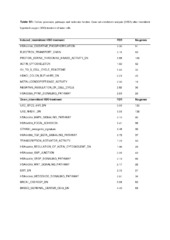
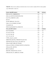
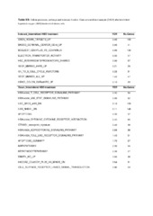
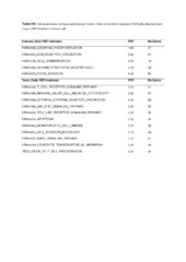
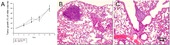
| dc.description.abstract | Background The tumor microenvironment is pivotal in tumor progression. Thus, we aimed to develop a mammary tumor model to elucidate molecular characteristics in the stroma versus the tumor cell compartment by global gene expression. Secondly, since tumor hypoxia influences several aspects of tumor pathophysiology, we hypothesized that hyperoxia might have an inhibitory effect on tumor growth per se. Finally, we aimed to identify differences in gene expression and key molecular mechanisms, both in the native state and following treatment. Methods 4T1 dsRed breast cancer cells were injected into eGFP expressing NOD/SCID mice. Group 1 was exposed to 3 intermittent HBO treatments (Day 1, 4 and 7), Group 2 to 7 daily HBO treatments (both 2.5bar, 100% O2, à 90 min), whereas the controls were exposed to a normal atmosphere. Tumor growth, histology, vascularisation, cell proliferation, cell death and metastasis were assessed. Fluorescence-activated cell sorting was used to separate tumor cells from stromal cells prior to gene expression analysis. Results The purity of sorted cells was verified by fluorescence microscopy. Gene expression profiling demonstrated that highly expressed genes in the untreated tumor stroma included constituents of the extracellular matrix and matrix metalloproteinases. Tumor growth was significantly inhibited by HBO, and the MAPK pathway was found to be significantly reduced. Immunohistochemistry indicated a significantly reduced microvessel density after intermittent HBO, whereas daily HBO did not show a similar effect. The anti-angiogenic response was reflected in the expression trends of angiogenic factors. Conclusions The present in vivo mammary tumor model enabled us to separate tumor and stromal cells, and demonstrated that the two compartments are characterized by distinct gene expressions, both in the native state and following HBO treatments. Furthermore, hyperoxia induced a significant tumor growth-inhibitory effect, with significant down-regulation of the MAPK pathway. An anti-angiogenic effect after intermittent HBO was observed, and reflected in the gene expression profile. | en_US |
| dc.language.iso | eng | eng |
| dc.publisher | BioMed Central Ltd. | eng |
| dc.rights | Attribution CC BY | eng |
| dc.rights.uri | http://creativecommons.org/licenses/by/2.0/ | eng |
| dc.title | Gene expression in tumor cells and stroma in dsRed 4T1 tumors in eGFP-expressing mice with and without enhanced oxygenation | en_US |
| dc.type | Peer reviewed | |
| dc.type | Journal article | |
| dc.description.version | publishedVersion | en_US |
| dc.rights.holder | Copyright 2012 Moen et al; licensee BioMed Central Ltd. | |
| dc.identifier.doi | https://doi.org/10.1186/1471-2407-12-21 | |
| dc.identifier.cristin | 922738 | |
| dc.source.journal | BMC Cancer | |
| dc.source.40 | 12 | |
| dc.source.14 | 21 |
Tilhørende fil(er)
Denne innførselen finnes i følgende samling(er)

Med mindre annet er angitt, så er denne innførselen lisensiert som Attribution CC BY