Cherenkov luminescence imaging for assessment of radioactive plaque position in brachytherapy of uveal melanoma: An in vivo feasibility study
| dc.contributor.author | Krohn, Jørgen Gitlesen | |
| dc.contributor.author | Chen, Yi-Chun | |
| dc.contributor.author | Stabo-Eeg, Nils Ole | |
| dc.contributor.author | Hamre, Børge | |
| dc.date.accessioned | 2021-02-12T10:54:20Z | |
| dc.date.available | 2021-02-12T10:54:20Z | |
| dc.date.created | 2020-09-17T12:07:50Z | |
| dc.date.issued | 2020 | |
| dc.Published | Translational Vision Science & Technology. 2020, (9) | en_US |
| dc.identifier.issn | 2164-2591 | |
| dc.identifier.uri | https://hdl.handle.net/11250/2727696 | |
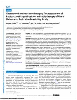
| dc.description.abstract | Purpose: To study the feasibility of using Cherenkov luminescence imaging (CLI) to evaluate and document ruthenium-106 plaque position during brachytherapy of uveal melanoma. Methods: Ruthenium-106 decays by emitting high-energy beta particles. When the electrons pass through the eye, Cherenkov radiation generates a faint light that can be captured by highly sensitive cameras. Patients undergoing ruthenium-106 plaque brachytherapy for posteriorly located choroidal melanoma were examined by CLI, which was performed in complete darkness with an electron multiplying charged-coupled device camera mounted on a fundus camera modified for long exposures. Results: Ten patients with tumors ranging from 5.8 to 13.0 mm in largest basal diameter and 2.0 to 4.6 mm in height were included. The plaques had an activity between 0.035 and 0.089 MBq/mm2 at the time of examination (1–4 days after implantation). CLI revealed the actual plaque position by displaying a circular area of light in the fundus corresponding with the plaque area. The Cherenkov light surrounded the tumor as a halo, which showed some asymmetry when the plaque was slightly displaced. The light intensity correlated positively with plaque activity and negatively with tumor pigmentation. Exposure times between 30 and 60 seconds were required to display the plaque position and delineate the tumor area. The long exposures made it difficult to maintain stable eye fixation and optimal image quality. Conclusions: CLI is a novel method to assess and document ruthenium-106 plaque position in brachytherapy for uveal melanoma. Translational Relevance: Ocular CLI may provide relevant radiation data during and after implantation of radioactive plaques, thus improving the accuracy of episcleral brachytherapy. | en_US |
| dc.language.iso | eng | en_US |
| dc.rights | Navngivelse 4.0 Internasjonal | * |
| dc.rights.uri | http://creativecommons.org/licenses/by/4.0/deed.no | * |
| dc.title | Cherenkov luminescence imaging for assessment of radioactive plaque position in brachytherapy of uveal melanoma: An in vivo feasibility study | en_US |
| dc.type | Journal article | en_US |
| dc.type | Peer reviewed | en_US |
| dc.description.version | publishedVersion | en_US |
| dc.rights.holder | Copyright 2020 The Authors | en_US |
| dc.source.articlenumber | 42 | en_US |
| cristin.ispublished | true | |
| cristin.fulltext | original | |
| cristin.qualitycode | 1 | |
| dc.identifier.doi | 10.1167/tvst.9.7.42 | |
| dc.identifier.cristin | 1830812 | |
| dc.source.journal | Translational Vision Science & Technology | en_US |
| dc.source.40 | 9 | en_US |
| dc.source.14 | 7 | en_US |
Tilhørende fil(er)
Denne innførselen finnes i følgende samling(er)
-
Department of Clinical Medicine [2044]
-
Registrations from Cristin [9489]

Med mindre annet er angitt, så er denne innførselen lisensiert som Navngivelse 4.0 Internasjonal