Liver Elastography in Primary Sclerosing Cholangitis Patients Using Three Different Scanner Systems
| dc.contributor.author | Mjelle, Anders Batman | |
| dc.contributor.author | Fossdal, Guri | |
| dc.contributor.author | Gilja, Odd Helge | |
| dc.contributor.author | Vesterhus, Mette | |
| dc.date.accessioned | 2021-03-03T11:17:51Z | |
| dc.date.available | 2021-03-03T11:17:51Z | |
| dc.date.created | 2020-11-17T09:15:41Z | |
| dc.date.issued | 2020 | |
| dc.Published | Ultrasound in Medicine and Biology. 2020, 46 (8), 1854-1864. | |
| dc.identifier.issn | 0301-5629 | |
| dc.identifier.uri | https://hdl.handle.net/11250/2731359 | |
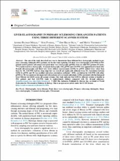
| dc.description.abstract | The aim of the study described here was to characterize three different liver elastography methods in primary sclerosing cholangitis (PSC) patients, for the first time exploring 2-D shear wave elastography (2-D-SWE) in PSC patients and its putative advantages over point shear wave elastography (pSWE). Sixty-six adult PSC patients (51 males, 77%) underwent liver elastography: Transient elastography (TE), pSWE and 2-D-SWE were applied head-to-head after B-mode ultrasonography and blood tests. Liver stiffness measurements (LSMs) by pSWE yielded lower values than those by TE; 2-D-SWE had less steep slope but was overall not significantly different from TE. Correlation between LSMs by pSWE and TE was excellent (intraclass correlation coefficient = 0.92); correlation for 2-D-SWE with either pSWE or TE was moderate but improved with exclusion of overweight individuals. LSMs correlated with the Enhanced Liver Fibrosis test (ELF) across all scanner systems. Our study indicates that LSM by different systems is feasible in PSC patients and that 2-D-SWE tends to underestimate stiffness compared with TE. | en_US |
| dc.language.iso | eng | en_US |
| dc.publisher | Elsevier | en_US |
| dc.rights | Navngivelse 4.0 Internasjonal | * |
| dc.rights.uri | http://creativecommons.org/licenses/by/4.0/deed.no | * |
| dc.title | Liver Elastography in Primary Sclerosing Cholangitis Patients Using Three Different Scanner Systems | en_US |
| dc.type | Journal article | en_US |
| dc.type | Peer reviewed | en_US |
| dc.description.version | publishedVersion | en_US |
| dc.rights.holder | Copyright 2020 2020 The Author(s). | en_US |
| cristin.ispublished | true | |
| cristin.fulltext | original | |
| cristin.qualitycode | 2 | |
| dc.identifier.doi | 10.1016/j.ultrasmedbio.2020.03.025 | |
| dc.identifier.cristin | 1848629 | |
| dc.source.journal | Ultrasound in Medicine and Biology | en_US |
| dc.source.40 | 46 | |
| dc.source.14 | 8 | |
| dc.source.pagenumber | 1854-1864 | en_US |
| dc.identifier.citation | Ultrasound in Medicine and Biology. 2020, 46 (8), 1854-1864 | en_US |
| dc.source.volume | 46 | en_US |
| dc.source.issue | 8 | en_US |
Tilhørende fil(er)
Denne innførselen finnes i følgende samling(er)
-
Department of Clinical Medicine [2044]
-
Registrations from Cristin [9489]

Med mindre annet er angitt, så er denne innførselen lisensiert som Navngivelse 4.0 Internasjonal