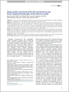
Image quality assessment of low-dose protocols in cone beam computed tomography of the anterior maxilla
| dc.contributor.author | Lynds, Randi | |
| dc.contributor.author | Kadesjö, Nils | |
| dc.contributor.author | Tsilingaridis, Georgios | |
| dc.contributor.author | Benchimol, Daniel | |
| dc.contributor.author | Shi, Xie Qi | |
| dc.date.accessioned | 2022-04-01T10:22:22Z | |
| dc.date.available | 2022-04-01T10:22:22Z | |
| dc.date.created | 2021-12-02T10:18:40Z | |
| dc.date.issued | 2022 | |
| dc.identifier.issn | 2212-4403 | |
| dc.identifier.uri | https://hdl.handle.net/11250/2989205 | |
| dc.description.abstract | Objectives. To evaluate overall image quality and visibility of anatomic structures on low-dose cone beam computed tomography (CBCT) scans and the effect of a noise reduction filter for assessment of the anterior maxilla. Methods. We obtained 48 CBCT volumes on 8 skull-phantoms using 6 protocols: 2 clinical default protocols [standard definition (SD) and high definition (HD)] and 4 low-dose protocols, 2 with a noise reduction filter [ultra-low-dose with high definition (ULDHD) and ultra-low-dose (ULD)] and 2 without [low-dose with high definition (LDHD) and low-dose (LD)]. Overall image quality and visibility of 8 anatomic structures were assessed by 5 observers and statistically analyzed using the Wilcoxon signed rank test. Intra- and interobserver agreement was measured using Cohen's weighted kappa. Results. HD provided higher overall image quality than diagnostically required; LD scored lower than diagnostically acceptable. ULDHD, ULD, and LDHD were acceptable. For anatomic structures, ULDHD and ULD were acceptable. LDHD and LD showed significantly inferior visibility for 1 and 4 structures, respectively. Mean values of intra- and interobserver agreement were 0.395 to 0.547 and 0.350 to 0.370, respectively. Conclusions. ULDHD, ULD, and LDHD may be recommended for assessment of impacted maxillary canines. The noise reduction filter affects image quality positively only at low exposure. | en_US |
| dc.language.iso | eng | en_US |
| dc.publisher | Elsevier | en_US |
| dc.rights | Navngivelse 4.0 Internasjonal | * |
| dc.rights.uri | http://creativecommons.org/licenses/by/4.0/deed.no | * |
| dc.title | Image quality assessment of low-dose protocols in cone beam computed tomography of the anterior maxilla | en_US |
| dc.type | Journal article | en_US |
| dc.type | Peer reviewed | en_US |
| dc.description.version | publishedVersion | en_US |
| dc.rights.holder | Copyright 2021 The Author(s) | en_US |
| cristin.ispublished | true | |
| cristin.fulltext | original | |
| cristin.qualitycode | 2 | |
| dc.identifier.doi | 10.1016/j.oooo.2021.10.001 | |
| dc.identifier.cristin | 1963174 | |
| dc.source.journal | Oral Surgery, Oral Medicine, Oral Pathology and Oral Radiology | en_US |
| dc.source.pagenumber | 483-491 | en_US |
| dc.identifier.citation | Oral Surgery, Oral Medicine, Oral Pathology and Oral Radiology. 2022, 133 (4), 483-491. | en_US |
| dc.source.volume | 133 | en_US |
| dc.source.issue | 4 | en_US |
Files in this item
This item appears in the following Collection(s)

Except where otherwise noted, this item's license is described as Navngivelse 4.0 Internasjonal