Human gingival epithelial cells stimulate proliferation, migration, and tube formation of lymphatic endothelial cells in vitro
| dc.contributor.author | Indrelid, Stine Hufthammer | |
| dc.contributor.author | Dongre, Harsh Nitin | |
| dc.contributor.author | Nunes, Ivana Pereira | |
| dc.contributor.author | Virtej, Anca | |
| dc.contributor.author | Bletsa, Athanasia | |
| dc.contributor.author | Berggreen, Ellen | |
| dc.date.accessioned | 2023-12-28T10:34:29Z | |
| dc.date.available | 2023-12-28T10:34:29Z | |
| dc.date.created | 2023-03-21T15:12:46Z | |
| dc.date.issued | 2023 | |
| dc.identifier.issn | 0022-3484 | |
| dc.identifier.uri | https://hdl.handle.net/11250/3108988 | |
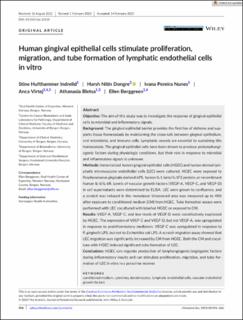
| dc.description.abstract | Objective The aim of this study was to investigate the response of gingival epithelial cells to microbial and inflammatory signals. Background The gingival epithelial barrier provides the first line of defense and supports tissue homeostasis by maintaining the cross-talk between gingival epithelium, oral microbiota, and immune cells. Lymphatic vessels are essential to sustaining this homeostasis. The gingival epithelial cells have been shown to produce prolymphangiogenic factors during physiologic conditions, but their role in response to microbial and inflammatory signals is unknown. Methods Immortalized human gingival epithelial cells (HGEC) and human dermal lymphatic microvascular endothelial cells (LEC) were cultured. HGEC were exposed to Porphyromonas gingivalis derived-LPS, human IL-1 beta/IL-1F2 protein, or recombinant human IL-6/IL-6R. Levels of vascular growth factors (VEGF-A, VEGF-C, and VEGF-D) in cell supernatants were determined by ELISA. LEC were grown to confluence, and a scratch was induced in the monolayer. Uncovered area was measured up to 48 h after exposure to conditioned medium (CM) from HGEC. Tube formation assays were performed with LEC cocultured with labelled HGEC or exposed to CM. Results VEGF-A, VEGF-C, and low levels of VEGF-D were constitutively expressed by HGEC. The expression of VEGF-C and VEGF-D, but not VEGF-A, was upregulated in response to proinflammatory mediators. VEGF-C was upregulated in response to P. gingivalis LPS, but not to Escherichia coli LPS. A scratch migration assay showed that LEC migration was significantly increased by CM from HGEC. Both the CM and coculture with HGEC induced significant tube formation of LEC. Conclusions HGEC can regulate production of lymphangiogenic/angiogenic factors during inflammatory insults and can stimulate proliferation, migration, and tube formation of LEC in vitro in a paracrine manner. | en_US |
| dc.language.iso | eng | en_US |
| dc.publisher | Wiley | en_US |
| dc.rights | Attribution-NonCommercial-NoDerivatives 4.0 Internasjonal | * |
| dc.rights.uri | http://creativecommons.org/licenses/by-nc-nd/4.0/deed.no | * |
| dc.title | Human gingival epithelial cells stimulate proliferation, migration, and tube formation of lymphatic endothelial cells in vitro | en_US |
| dc.type | Journal article | en_US |
| dc.type | Peer reviewed | en_US |
| dc.description.version | publishedVersion | en_US |
| dc.rights.holder | Copyright 2023 The Author(s) | en_US |
| cristin.ispublished | true | |
| cristin.fulltext | original | |
| cristin.qualitycode | 1 | |
| dc.identifier.doi | 10.1111/jre.13110 | |
| dc.identifier.cristin | 2135871 | |
| dc.source.journal | Journal of Periodontal Research | en_US |
| dc.source.pagenumber | 596-606 | en_US |
| dc.identifier.citation | Journal of Periodontal Research. 2023, 58 (3), 596-606. | en_US |
| dc.source.volume | 58 | en_US |
| dc.source.issue | 3 | en_US |
Tilhørende fil(er)
Denne innførselen finnes i følgende samling(er)

Med mindre annet er angitt, så er denne innførselen lisensiert som Attribution-NonCommercial-NoDerivatives 4.0 Internasjonal