| dc.contributor.author | Pålhaugen, Lene | |
| dc.contributor.author | Sudre, Carole H | |
| dc.contributor.author | Teceläo, Sandra Raquel Ramos | |
| dc.contributor.author | Nakling, Arne | |
| dc.contributor.author | Almdahl, Ina Selseth | |
| dc.contributor.author | Kalheim, Lisa Flem | |
| dc.contributor.author | Cardoso, M Jorge | |
| dc.contributor.author | Johnsen, Stein Harald | |
| dc.contributor.author | Rongve, Arvid | |
| dc.contributor.author | Aarsland, Dag | |
| dc.contributor.author | Bjørnerud, Atle | |
| dc.contributor.author | Selnes, Per | |
| dc.contributor.author | Fladby, Tormod | |
| dc.date.accessioned | 2021-04-30T09:39:10Z | |
| dc.date.available | 2021-04-30T09:39:10Z | |
| dc.date.created | 2020-09-28T09:07:17Z | |
| dc.date.issued | 2020 | |
| dc.Published | Journal of Cerebral Blood Flow and Metabolism. 2020, 1-13. | |
| dc.identifier.issn | 0271-678X | |
| dc.identifier.uri | https://hdl.handle.net/11250/2740555 | |
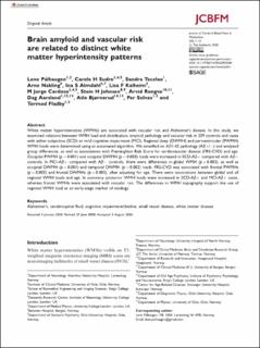
| dc.description.abstract | White matter hyperintensities (WMHs) are associated with vascular risk and Alzheimer’s disease. In this study, we examined relations between WMH load and distribution, amyloid pathology and vascular risk in 339 controls and cases with either subjective (SCD) or mild cognitive impairment (MCI). Regional deep (DWMH) and periventricular (PWMH) WMH loads were determined using an automated algorithm. We stratified on Aβ1-42 pathology (Aβ+/−) and analyzed group differences, as well as associations with Framingham Risk Score for cardiovascular disease (FRS-CVD) and age. Occipital PWMH (p = 0.001) and occipital DWMH (p = 0.003) loads were increased in SCD-Aβ+ compared with Aβ− controls. In MCI-Aβ+ compared with Aβ− controls, there were differences in global WMH (p = 0.003), as well as occipital DWMH (p = 0.001) and temporal DWMH (p = 0.002) loads. FRS-CVD was associated with frontal PWMHs (p = 0.003) and frontal DWMHs (p = 0.005), after adjusting for age. There were associations between global and all regional WMH loads and age. In summary, posterior WMH loads were increased in SCD-Aβ+ and MCI-Aβ+ cases, whereas frontal WMHs were associated with vascular risk. The differences in WMH topography support the use of regional WMH load as an early-stage marker of etiology. | en_US |
| dc.language.iso | eng | en_US |
| dc.publisher | Sage | en_US |
| dc.rights | Navngivelse 4.0 Internasjonal | * |
| dc.rights.uri | http://creativecommons.org/licenses/by/4.0/deed.no | * |
| dc.title | Brain amyloid and vascular risk are related to distinct white matter hyperintensity patterns | en_US |
| dc.type | Journal article | en_US |
| dc.type | Peer reviewed | en_US |
| dc.description.version | publishedVersion | en_US |
| dc.rights.holder | Copyright 2020 The Authors | en_US |
| cristin.ispublished | true | |
| cristin.fulltext | original | |
| cristin.qualitycode | 2 | |
| dc.identifier.doi | 10.1177/0271678X20957604 | |
| dc.identifier.cristin | 1833921 | |
| dc.source.journal | Journal of Cerebral Blood Flow and Metabolism | en_US |
| dc.source.pagenumber | 1162-1174 | en_US |
| dc.relation.project | Norges forskningsråd: 269774 | en_US |
| dc.identifier.citation | Journal of Cerebral Blood Flow and Metabolism. 2020, 41(5):1162-1174 | en_US |
| dc.source.volume | 41 | en_US |
| dc.source.issue | 5 | en_US |

