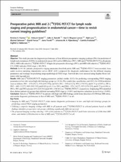
Preoperative pelvic MRI and 2-[18F]FDG PET/CT for lymph node staging and prognostication in endometrial cancer—time to revisit current imaging guidelines?
| dc.contributor.author | Fasmer, Kristine Eldevik | |
| dc.contributor.author | Gulati, Ankush | |
| dc.contributor.author | Dybvik, Julie Andrea | |
| dc.contributor.author | Wagner-Larsen, Kari Strøno | |
| dc.contributor.author | Lura, Njål Gjærde | |
| dc.contributor.author | Salvesen, Øyvind | |
| dc.contributor.author | Forsse, David Erik | |
| dc.contributor.author | Trovik, Jone | |
| dc.contributor.author | Pijnenborg, Johanna M. A. | |
| dc.contributor.author | Krakstad, Camilla | |
| dc.contributor.author | Haldorsen, Ingfrid S. | |
| dc.date.accessioned | 2022-09-27T12:19:42Z | |
| dc.date.available | 2022-09-27T12:19:42Z | |
| dc.date.created | 2022-09-26T10:36:52Z | |
| dc.date.issued | 2022 | |
| dc.identifier.issn | 0938-7994 | |
| dc.identifier.uri | https://hdl.handle.net/11250/3021797 | |
| dc.description.abstract | Objective This study presents the diagnostic performance of four different preoperative imaging workups (IWs) for prediction of lymph node metastases (LNMs) in endometrial cancer (EC): pelvic MRI alone (IW1), MRI and [18F]FDG-PET/CT in all patients (IW2), MRI with selective [18F]FDG-PET/CT if high-risk preoperative histology (IW3), and MRI with selective [18F]FDG-PET/CT if MRI indicates FIGO stage ≥ 1B (IW4). Methods In 361 EC patients, preoperative staging parameters from both pelvic MRI and [18F]FDG-PET/CT were recorded. Area under receiver operating characteristic curves (ROC AUC) compared the diagnostic performance for the different imaging parameters and workups for predicting surgicopathological FIGO stage. Survival data were assessed using Kaplan-Meier estimator with log-rank test. Results MRI and [18F]FDG-PET/CT staging parameters yielded similar AUCs for predicting corresponding FIGO staging parameters in low-risk versus high-risk histology groups (p ≥ 0.16). The sensitivities, specificities, and AUCs for LNM prediction were as follows: IW1—33% [9/27], 95% [185/193], and 0.64; IW2—56% [15/27], 90% [174/193], and 0.73 (p = 0.04 vs. IW1); IW3—44% [12/27], 94% [181/193], and 0.69 (p = 0.13 vs. IW1); and IW4—52% [14/27], 91% [176/193], and 0.72 (p = 0.06 vs. IW1). IW3 and IW4 selected 34% [121/361] and 54% [194/361] to [18F]FDG-PET/CT, respectively. Employing IW4 identified three distinct patient risk groups that exhibited increasing FIGO stage (p < 0.001) and stepwise reductions in survival (p ≤ 0.002). Conclusion Selective [18F]FDG-PET/CT in patients with high-risk MRI findings yields better detection of LNM than MRI alone, and similar diagnostic performance to that of MRI and [18F]FDG-PET/CT in all. | en_US |
| dc.language.iso | eng | en_US |
| dc.publisher | Springer | en_US |
| dc.rights | Navngivelse 4.0 Internasjonal | * |
| dc.rights.uri | http://creativecommons.org/licenses/by/4.0/deed.no | * |
| dc.title | Preoperative pelvic MRI and 2-[18F]FDG PET/CT for lymph node staging and prognostication in endometrial cancer—time to revisit current imaging guidelines? | en_US |
| dc.type | Journal article | en_US |
| dc.type | Peer reviewed | en_US |
| dc.description.version | publishedVersion | en_US |
| dc.rights.holder | Copyright The Author(s) 2022 | en_US |
| cristin.ispublished | true | |
| cristin.fulltext | original | |
| cristin.qualitycode | 2 | |
| dc.identifier.doi | 10.1007/s00330-022-08949-3 | |
| dc.identifier.cristin | 2055307 | |
| dc.source.journal | European Radiology | en_US |
| dc.identifier.citation | European Radiology, 2022. | en_US |
Tilhørende fil(er)
Denne innførselen finnes i følgende samling(er)
-
Department of Clinical Medicine [2043]
-
Registrations from Cristin [9482]

Med mindre annet er angitt, så er denne innførselen lisensiert som Navngivelse 4.0 Internasjonal