| dc.contributor.author | Nylund, Are | |
| dc.contributor.author | Hansen, Haakon | |
| dc.contributor.author | Brevik, Øyvind Jakobsen | |
| dc.contributor.author | Hustoft, Håvard | |
| dc.contributor.author | Markussen, Turhan | |
| dc.contributor.author | Plarre, Heidrun | |
| dc.contributor.author | Karlsbakk, Egil | |
| dc.date.accessioned | 2019-02-13T14:52:16Z | |
| dc.date.available | 2019-02-13T14:52:16Z | |
| dc.date.issued | 2018-01-06 | |
| dc.Published | Nylund A, Hansen H, Brevik ØJ, Hustoft H, Markussen T, Plarre H, Karlsbakk E. Infection dynamics and tissue tropism of Parvicapsula pseudobranchicola (Myxozoa: Myxosporea) in farmed Atlantic salmon (Salmo salar). Parasites & Vectors. 2018;11:17:1-13 | eng |
| dc.identifier.issn | 1756-3305 | en_US |
| dc.identifier.uri | https://hdl.handle.net/1956/19097 | |
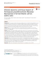
| dc.description.abstract | Background The myxosporean parasite Parvicapsula pseudobranchicola commonly infects farmed Atlantic salmon in northern Norway. Heavy infections are associated with pseudobranch lesions, runting and mortality in the salmon populations. The life-cycle of the parasite is unknown, preventing controlled challenge experiments. The infection dynamics, duration of sporogony, tissue tropism and ability to develop immunity to the parasite in farmed Atlantic salmon is poorly known. We conducted a field experiment, aiming at examining these aspects. Methods Infections in a group of Atlantic salmon were followed from before sea-transfer to the end of the production (604 days). Samples from a range of tissues/sites were analysed using real-time RT-PCR and histology, including in situ hybridization. Results All salmon in the studied population rapidly became infected with P. pseudobranchicola after sea-transfer medio August. Parasite densities in the pseudobranchs peaked in winter (November-January), and decreased markedly to March. Densities thereafter decreased further. Parasite densities in other tissues were low. Parasite stages were initially found to be intravascular in the pseudobranch, but occurred extravascular in the pseudobranch tissue at 3 months post-sea-transfer. Mature spores appeared in the pseudobranchs in the period with high parasite densities in the winter (late November-January), and were released (i.e. disappeared from the fish) in the period January-March. Clinical signs of parvicapsulosis (December-early February) were associated with high parasite densities and inflammation in the pseudobranchs. No evidence for reinfection was seen the second autumn in sea. Conclusions The main site of the parasite in Atlantic salmon is the pseudobranchs. Blood stages occur, but parasite proliferation is primarily associated with extravascular stages in the pseudobranchs. Disease and mortality (parvicapsulosis) coincide with the completion of sporogony. Atlantic salmon appears to develop immunity to P. pseudobranchicola. Further studies should focus on the unknown life-cycle of the parasite, and the pathophysiological effects of the pseudobranch infection that also could affect the eyes and vision. | en_US |
| dc.language.iso | eng | eng |
| dc.publisher | Springer Nature | en_US |
| dc.rights | Attribution CC BY | eng |
| dc.rights.uri | http://creativecommons.org/licenses/by/4.0/ | eng |
| dc.subject | Parasites | eng |
| dc.subject | Salmo Salar | eng |
| dc.subject | Aquaculture | eng |
| dc.subject | Disease | eng |
| dc.subject | Norway | eng |
| dc.subject | in situ hybridization | eng |
| dc.subject | Real-time RT-PCR | eng |
| dc.title | Infection dynamics and tissue tropism of Parvicapsula pseudobranchicola (Myxozoa: Myxosporea) in farmed Atlantic salmon (Salmo salar) | en_US |
| dc.type | Peer reviewed | |
| dc.type | Journal article | |
| dc.date.updated | 2018-07-09T09:11:44Z | |
| dc.description.version | publishedVersion | en_US |
| dc.rights.holder | Copyright The Author(s) 2018 | en_US |
| dc.identifier.doi | https://doi.org/10.1186/s13071-017-2583-9 | |
| dc.identifier.cristin | 1589034 | |
| dc.source.journal | Parasites & Vectors | |

