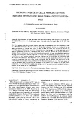
Microfilaments in cells associated with induced heterotpic bone formation in guinea pigs
Journal article
Permanent lenke
https://hdl.handle.net/1956/2188Utgivelsesdato
1980Metadata
Vis full innførselSamlinger
Sammendrag
Sera from active chronic hepatitis were used to demonstrate actin microfilaments in cells of a bone-induced model. Demnineralized and freeze-dried allogenic dentine was implanted in the abdominal muscles of Guinea pigs. After 21 and 28 days the tissue was excised and showed: a) resorption areas of the dentine contained dentinoclasts and macrophages. B) osteoid and bone and c) areas of fibroblastic proliferation. Osteoblasts and young osteocytes showed the strongest fluorescent staining. Electron microscopy of these cells demonstrated abundant 5-7 nm microfilaments interpreted as actin in the outer parts and cell projections, in which some microtubules also were observed. Fewer microfilaments were present in mature osteocytes although the cell projections in mineralized bone also showed microfilaments. Microfilaments were also found in some dentinoclasts where the actin was located in projections from the ruffles border protruding into the dentine. The localization in the ruffled border corresponded to the distribution of the fluorescent staining of actin. The high amount of actin in osteoblasts and osteocytes may be correlated to the synthesis and transport of the new matrix components. The microfilaments in the clear sealing zone of dentinoclasts were considered to have a function in the sealing of the ruffled border.