Altered development of fetal liver perfusion in pregnancies with pregestational diabetes
| dc.contributor.author | Lund, Agnethe | en_US |
| dc.contributor.author | Ebbing, Cathrine | en_US |
| dc.contributor.author | Rasmussen, Svein | en_US |
| dc.contributor.author | Kiserud, Torvid | en_US |
| dc.contributor.author | Hanson, Mark | en_US |
| dc.contributor.author | Kessler, Jørg | en_US |
| dc.date.accessioned | 2020-05-12T12:43:40Z | |
| dc.date.available | 2020-05-12T12:43:40Z | |
| dc.date.issued | 2019-03-13 | |
| dc.Published | Lund A, Ebbing C, Rasmussen S, Kiserud T, Hanson M, Kessler J. Altered development of fetal liver perfusion in pregnancies with pregestational diabetes. PLOS ONE. 2019;14(3):e0211788 | eng |
| dc.identifier.issn | 1932-6203 | |
| dc.identifier.uri | https://hdl.handle.net/1956/22205 | |
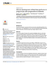
| dc.description.abstract | Background: Pregestational diabetes is associated with fetal macrosomia, and umbilical perfusion of the fetal liver has a role in regulating fetal growth. We therefore hypothesized that pregestational diabetes alters fetal liver blood flow depending on degree of glycemic control. Methods: In a prospective study, 49 women with pregestational diabetes underwent monthly ultrasound examinations during 24–36 gestational weeks. Blood flow was determined in the umbilical vein, ductus venosus and portal vein, and blood velocity was measured in the left portal vein, the latter reflecting the watershed between splanchnic and umbilical flow. The measurements were compared with reference values by z-score statistics, and the effect of HbA1c assessed. Results: The umbilical venous flow to the liver (z-score 0.36, p = 0.002), total venous liver flow (z-score 0.51, p<0.001) and left portal vein blood velocity (z-score 0.64, p<0.001), were higher in the study group. Normalized portal venous flow was lower (z-score -0.42, p = 0.002), and normalized total venous liver flow tended to be lower after 30 gestational weeks (z-score -0.54, p = 0.047) in the diabetic pregnancies compared with reference values from a low-risk population. The left portal vein blood velocity was positively, and the portal fraction of total venous liver flow negatively correlated with first trimester HbA1C. Conclusions: In spite of increased umbilical blood distribution to the fetal liver, graded according to glycemic control, the total venous liver flow did not match third trimester fetal growth in pregnancies with pregestational diabetes, thus contributing towards increased perinatal risks and possibly altered liver function with long-term metabolic consequences. | en_US |
| dc.language.iso | eng | eng |
| dc.publisher | PLOS | eng |
| dc.rights | Attribution CC BY | eng |
| dc.rights.uri | http://creativecommons.org/licenses/by/4.0 | eng |
| dc.title | Altered development of fetal liver perfusion in pregnancies with pregestational diabetes | en_US |
| dc.type | Peer reviewed | |
| dc.type | Journal article | |
| dc.date.updated | 2019-12-04T14:58:21Z | |
| dc.description.version | publishedVersion | en_US |
| dc.rights.holder | Copyright 2019 Lund et al. | |
| dc.identifier.doi | https://doi.org/10.1371/journal.pone.0211788 | |
| dc.identifier.cristin | 1714262 | |
| dc.source.journal | PLoS ONE |
Tilhørende fil(er)
Denne innførselen finnes i følgende samling(er)

Med mindre annet er angitt, så er denne innførselen lisensiert som Attribution CC BY