Endorectal ultrasonography, strain elastography and MRI differentiation of rectal adenomas and adenocarcinomas.
| dc.contributor.author | Waage, Jo Erling Riise | en_US |
| dc.contributor.author | Leh, Sabine Maria | en_US |
| dc.contributor.author | Røsler, Cornelia | en_US |
| dc.contributor.author | Pfeffer, Frank | en_US |
| dc.contributor.author | Bach, Simon | en_US |
| dc.contributor.author | Havre, Roald Flesland | en_US |
| dc.contributor.author | Haldorsen, Ingfrid S. | en_US |
| dc.contributor.author | Ødegaard, Svein | en_US |
| dc.contributor.author | Baatrup, Gunnar | en_US |
| dc.date.accessioned | 2015-03-25T13:52:17Z | |
| dc.date.available | 2015-03-25T13:52:17Z | |
| dc.date.issued | 2015-02 | eng |
| dc.identifier.issn | 1462-8910 | |
| dc.identifier.uri | https://hdl.handle.net/1956/9655 | |
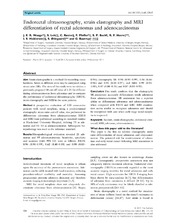
| dc.description.abstract | Aim Strain elastography is a method for recording tissue hardness. Strain in different areas may be compared using strain ratio (SR). The aims of this study were to validate a previously proposed SR cut-off value of 1.25 for differentiating adenocarcinomas from adenomas and to compare the performance of endorectal ultrasonography (ERUS), strain elastography and MRI in the same patients. Method A prospective evaluation of 120 consecutive patients with rectal neoplasia, using a predetermined elastography strain ratio cut-off value, was performed to differentiate adenomas from adenocarcinomas. ERUS and MRI were performed according to standard routine at Haukeland University Hospital, defining T0 as adenomas and T1–T4 as adenocarcinomas. Subsequent histopathology was used as the reference standard. Results Histopathological evaluation revealed 21 adenomas and 99 adenocarcinomas. Sensitivity, specificity and accuracy (with 95% CI) were as follows: ERUS: 0.96 (0.90–0.99), 0.62 (0.40–0.80) and 0.90 (0.83–0.94); elastography SR: 0.96 (0.90–0.99), 0.86 (0.66–0.96) and 0.94 (0.88–0.97); and MRI: 0.99 (0.94–1.00), 0.07 (0.00–0.31) and 0.87 (0.80–0.93). Conclusion This study confirms that the elastography SR assessment accurately differentiates sessile adenomas from adenocarcinomas. SR assessment has a superior ability to differentiate adenomas and adenocarcinomas when compared with ERUS and MRI. MRI examination seems unable to recognize adenomas and should be interpreted with care when early-stage rectal neoplasia is suspected. | en_US |
| dc.language.iso | eng | eng |
| dc.publisher | Wiley | eng |
| dc.rights | Attribution-NonCommercial-NoDerivs CC BY-NC-ND | eng |
| dc.rights.uri | http://creativecommons.org/licenses/by-nc-nd/4.0/ | eng |
| dc.subject | Rectum | eng |
| dc.subject | strain elastography | eng |
| dc.subject | endorectal ultrasound | eng |
| dc.subject | MRI | eng |
| dc.subject | adenoma | eng |
| dc.subject | adenocarcinoma | eng |
| dc.title | Endorectal ultrasonography, strain elastography and MRI differentiation of rectal adenomas and adenocarcinomas. | en_US |
| dc.type | Peer reviewed | |
| dc.type | Journal article | |
| dc.date.updated | 2015-03-03T10:02:44Z | en_US |
| dc.description.version | publishedVersion | en_US |
| dc.rights.holder | Copyright 2014 The Authors | |
| dc.identifier.doi | https://doi.org/10.1111/codi.12845 | |
| dc.identifier.cristin | 1221747 | |
| dc.source.journal | Colorectal Disease | |
| dc.source.40 | 17 | |
| dc.source.14 | 2 | |
| dc.source.pagenumber | 124-131 |
Files in this item
This item appears in the following Collection(s)

Except where otherwise noted, this item's license is described as Attribution-NonCommercial-NoDerivs CC BY-NC-ND