Ciliary photoreceptors in the cerebral eyes of a protostome larva
| dc.contributor.author | Passamaneck, Yale J. | eng |
| dc.contributor.author | Furchheim, Nina | eng |
| dc.contributor.author | Hejnol, Andreas | eng |
| dc.contributor.author | Martindale, Mark Q. | eng |
| dc.contributor.author | Lüter, Carsten | eng |
| dc.date.accessioned | 2012-01-24T11:15:10Z | |
| dc.date.available | 2012-01-24T11:15:10Z | |
| dc.date.issued | 2011-03-01 | eng |
| dc.Published | EvoDevo 2011, 2:6 | en |
| dc.identifier.issn | 2041-9139 | |
| dc.identifier.uri | https://hdl.handle.net/1956/5520 | |
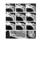
| dc.description.abstract | Background: Eyes in bilaterian metazoans have been described as being composed of either ciliary or rhabdomeric photoreceptors. Phylogenetic distribution, as well as distinct morphologies and characteristic deployment of different photopigments (ciliary vs. rhabdomeric opsins) and transduction pathways argue for the co-existence of both of these two photoreceptor types in the last common bilaterian ancestor. Both receptor types exist throughout the Bilateria, but only vertebrates are thought to use ciliary photoreceptors for directional light detection in cerebral eyes, while all other invertebrate bilaterians studied utilize rhabdomeric photoreceptors for this purpose. In protostomes, ciliary photoreceptors that express c-opsin have been described only from a nonvisual deep-brain photoreceptor. Their homology with vertebrate rods and cones of the human eye has been hypothesized to represent a unique functional transition from non-visual to visual roles in the vertebrate lineage. Results: To test the hypothesis that protostome cerebral eyes employ exclusively rhabdomeric photoreceptors, we investigated the ultrastructure of the larval eyes in the brachiopod Terebratalia transversa. We show that these pigment-cup eyes consist of a lens cell and a shading pigment cell, both of which are putative photoreceptors, deploying a modified, enlarged cilium for light perception, and have axonal connections to the larval brain. Our investigation of the gene expression patterns of c-opsin, Pax6 and otx in these eyes confirms that the larval eye spots of brachiopods are cerebral eyes that deploy ciliary type photoreceptors for directional light detection. Interestingly, c-opsin is also expressed during early embryogenesis in all potential apical neural cells, becoming restricted to the anterior neuroectoderm, before expression is initiated in the photoreceptor cells of the eyes. Coincident with the expression of c-opsin in the presumptive neuroectoderm, we found that middle gastrula stage embryos display a positive photoresponse behavior, in the absence of a discrete shading pigment or axonal connections between cells. Conclusions: Our results indicate that the dichotomy in the deployment of ciliary and rhabdomeric photoreceptors for directional light detection is not as clear-cut as previously thought. Analyses of brachiopod larval eyes demonstrate that the utilization of c-opsin expressing ciliary photoreceptors in cerebral eyes is not limited to vertebrates. The presence of ciliary photoreceptor-based eyes in protostomes suggests that the transition between non-visual and visual functions of photoreceptors has been more evolutionarily labile than previously recognized, and that co-option of ciliary and rhabdomeric photoreceptor cell types for directional light detection has occurred multiple times during animal evolution. In addition, positive photoresponse behavior in gastrula stage embryos suggests that a discrete shading pigment is not requisite for directional photoreception in metazoans. Scanning photoreception of light intensities mediating cell-autonomous changes of ciliary movement may represent an ancient mechanism for regulating locomotory behavior, and is likely to have existed prior to the evolution of eye-mediated directional light detection employing axonal connections to effector cells and a discreet shading pigment. | en_US |
| dc.language.iso | eng | eng |
| dc.publisher | BioMed Central | eng |
| dc.rights | Attribution CC BY | eng |
| dc.rights.uri | http://creativecommons.org/licenses/by/2.0/ | eng |
| dc.title | Ciliary photoreceptors in the cerebral eyes of a protostome larva | eng |
| dc.type | Peer reviewed | eng |
| dc.type | Journal article | eng |
| dc.description.version | publishedVersion | |
| dc.rights.holder | Copyright 2011 Passamaneck et al; licensee BioMed Central Ltd. | |
| dc.identifier.doi | https://doi.org/10.1186/2041-9139-2-6 | |
| dc.identifier.cristin | 1042470 | |
| dc.subject.nsi | VDP::Mathematics and natural science: 400::Zoology and botany: 480::Marine biology: 497 | eng |
| dc.subject.nsi | VDP::Mathematics and natural science: 400::Basic biosciences: 470::General microbiology: 472 | eng |
Tilhørende fil(er)

Denne innførselen finnes i følgende samling(er)

Med mindre annet er angitt, så er denne innførselen lisensiert som Attribution CC BY