Threshold estimation based on local minima for nucleus and cytoplasm segmentation
| dc.contributor.author | Mayala, Simeon Sahani | |
| dc.contributor.author | Haugsøen, Jonas Bull | |
| dc.date.accessioned | 2022-08-10T07:12:45Z | |
| dc.date.available | 2022-08-10T07:12:45Z | |
| dc.date.created | 2022-04-26T16:43:02Z | |
| dc.date.issued | 2022 | |
| dc.identifier.issn | 1471-2342 | |
| dc.identifier.uri | https://hdl.handle.net/11250/3010963 | |
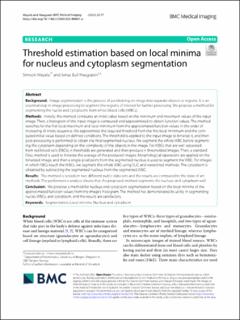
| dc.description.abstract | Background: Image segmentation is the process of partitioning an image into separate objects or regions. It is an essential step in image processing to segment the regions of interest for further processing. We propose a method for segmenting the nuclei and cytoplasms from white blood cells (WBCs). Methods: Initially, the method computes an initial value based on the minimum and maximum values of the input image. Then, a histogram of the input image is computed and approximated to obtain function values. The method searches for the first local maximum and local minimum from the approximated function values in the order of increasing of knots sequence. We approximate the required threshold from the first local minimum and the computed initial value based on defined conditions. The threshold is applied to the input image to binarize it, and then post-processing is performed to obtain the final segmented nucleus. We segment the whole WBC before segmenting the cytoplasm depending on the complexity of the objects in the image. For WBCs that are well separated from red blood cells (RBCs), n thresholds are generated and then produce n thresholded images. Then, a standard Otsu method is used to binarize the average of the produced images. Morphological operations are applied on the binarized image, and then a single-pixel point from the segmented nucleus is used to segment the WBC. For images in which RBCs touch the WBCs, we segment the whole WBC using SLIC and watershed methods. The cytoplasm is obtained by subtracting the segmented nucleus from the segmented WBC. Results: The method is tested on two different public data sets and the results are compared to the state of art methods. The performance analysis shows that the proposed method segments the nucleus and cytoplasm well. Conclusion: We propose a method for nucleus and cytoplasm segmentation based on the local minima of the approximated function values from the image’s histogram. The method has demonstrated its utility in segmenting nuclei, WBCs, and cytoplasm, and the results are satisfactory. | en_US |
| dc.language.iso | eng | en_US |
| dc.publisher | BMC | en_US |
| dc.rights | Navngivelse 4.0 Internasjonal | * |
| dc.rights.uri | http://creativecommons.org/licenses/by/4.0/deed.no | * |
| dc.title | Threshold estimation based on local minima for nucleus and cytoplasm segmentation | en_US |
| dc.type | Journal article | en_US |
| dc.type | Peer reviewed | en_US |
| dc.description.version | publishedVersion | en_US |
| dc.rights.holder | Copyright The Author(s) 2022 | en_US |
| dc.source.articlenumber | 77 | en_US |
| cristin.ispublished | true | |
| cristin.fulltext | original | |
| cristin.qualitycode | 1 | |
| dc.identifier.doi | 10.1186/s12880-022-00801-w | |
| dc.identifier.cristin | 2019290 | |
| dc.source.journal | BMC Medical Imaging | en_US |
| dc.identifier.citation | BMC Medical Imaging. 2022, 22, 77. | en_US |
| dc.source.volume | 22 | en_US |
Tilhørende fil(er)
Denne innførselen finnes i følgende samling(er)
-
Department of Mathematics [939]
-
Registrations from Cristin [9791]

Med mindre annet er angitt, så er denne innførselen lisensiert som Navngivelse 4.0 Internasjonal