| dc.contributor.author | Parkar, Anagha P | |
| dc.contributor.author | Adriaensen, Miraude E.A.M.P. | |
| dc.contributor.author | Vindfeld, Søren | |
| dc.contributor.author | Solheim, Eirik | |
| dc.date.accessioned | 2021-09-14T12:29:19Z | |
| dc.date.available | 2021-09-14T12:29:19Z | |
| dc.date.created | 2016-12-18T19:28:17Z | |
| dc.date.issued | 2016 | |
| dc.identifier.issn | 0363-5465 | |
| dc.identifier.uri | https://hdl.handle.net/11250/2776504 | |
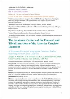
| dc.description.abstract | Background:
The anterior cruciate ligament (ACL) is regularly reconstructed if knee joint function is impaired. Anatomic graft tunnel placement, often assessed with varying measurement methods, in the femur and tibia is considered important for an optimal clinical outcome. A consensus on the exact location of the femoral and tibial footprint centers is lacking.
Purpose:
To systematically review the literature regarding anatomic centers of the femoral and tibial ACL footprints and assess the mean, median, and percentiles of normal centers.
Study Design:
Systematic review.
Methods:
A systematic literature search was performed in the PubMed/Medline database in November 2015. Search terms were the following: “ACL” and “insertion anatomy” or “anatomic footprint” or “radiographic landmarks” or “quadrant methods” or “tunnel placement” or “cadaveric femoral” or “cadaveric tibial.” English-language articles that reported the location of the ACL footprint according to the Bernard and Hertel grid in the femur and the Stäubli and Rauschning method in the tibia were included. Weighted means, weighted medians, and weighted 5th and 95th percentiles were calculated.
Results:
The initial search yielded 1393 articles. After applying the inclusion and exclusion criteria, 16 studies with measurements on cadaveric specimens or a healthy population were reviewed. The weighted mean of the femoral insertion center based on measurements in 218 knees was 29% in the deep-shallow (DS) direction and 35% in the high-low (HL) direction. The weighted median was 26% for DS and 34% for HL. The weighted 5th and 95th percentiles for DS were 24% and 37%, respectively, and for HL were 28% and 43%, respectively. The weighted mean of the tibial insertion center in the anterior-posterior direction based on measurements in 300 knees was 42%, and the weighted median was 44%; the 5th and 95th percentiles were 39% and 46%, respectively.
Conclusion:
Our results show slight differences between the weighted means and medians in the femoral and tibial insertion centers. We recommend the use of the 5th and 95th percentiles when considering postoperative placement to be “in or out of the anatomic range.” | en_US |
| dc.language.iso | eng | en_US |
| dc.publisher | Sage | en_US |
| dc.title | The anatomic centers of the femoral and tibial insertions of the anterior cruciate ligament: a systematic review of imaging and cadaveric studies reporting normal center locations | en_US |
| dc.type | Journal article | en_US |
| dc.type | Peer reviewed | en_US |
| dc.description.version | acceptedVersion | en_US |
| dc.rights.holder | Copyright 2016 The Author(s) | en_US |
| cristin.ispublished | true | |
| cristin.fulltext | postprint | |
| cristin.qualitycode | 2 | |
| dc.identifier.doi | 10.1177/0363546516673984 | |
| dc.identifier.cristin | 1414731 | |
| dc.source.journal | American Journal of Sports Medicine | en_US |
| dc.source.pagenumber | 2180-2188 | en_US |
| dc.identifier.citation | American Journal of Sports Medicine. 2016, 45 (9), 2180-2188. | en_US |
| dc.source.volume | 45 | en_US |
| dc.source.issue | 9 | en_US |
