Metastatic tumor of the interventricular septum mimicking myocardial calcification: The role of multimodality imaging
Journal article, Peer reviewed
Published version

Åpne
Permanent lenke
https://hdl.handle.net/11250/2833497Utgivelsesdato
2021Metadata
Vis full innførselSamlinger
- Department of Biomedicine [742]
- Registrations from Cristin [10412]
Sammendrag
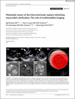
Recently, there has been focus on how a round-shaped metastatic or dystrophic myocardial calcification, a rare type of myocardial pathology with different imaging appearances, can mimic a tumor in the basal interventricular septum (IVS)1 on transthoracic echocardiography. However, we should also be focused on myocardial tumors without calcification. Here, we present a case of a 67-year-old woman with a tumor in the basal IVS detected by a routine echocardiography. She was previously diagnosed with a metastatic leiomyosarcoma, but did not have a known cardiovascular disorder. She did not have history of coronary artery disease, diabetes, or hypertension, and her blood pressure values at outpatient clinic were within normal range (<140/90 mmHg). A well-defined, round-shaped tumor (26 × 23 mm) was seen in the basal IVS (Supplementary data online, Videos 1 and 2) that was not present on an echocardiogram only 16 months earlier (Figure 1A), but evident t follow-up (Figure 1B-C). There was no signs of left ventricular hypertrophy or significant valvular heart disease. For better tissue characterization and assessment of the atrial septum and adjacent epicardium, a cardiac magnetic resonance (CMR) was performed (Figure 1E–G), confirming findings observed on echocardiography. In addition, it also identified two other tumors, one in the atrial septum (Figure 1G, white arrow) and another in the pericardium (Figure 1G, red arrow), highlighting the importance of multimodality imaging.
