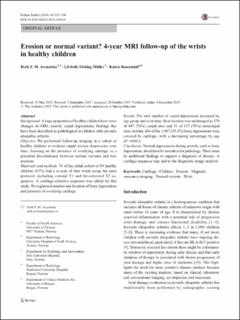
Erosion or normal variant? 4-year MRI follow-up of the wrists in healthy children
| dc.contributor.author | Avenarius, Derk | |
| dc.contributor.author | Müller, Lil-Sofie Ording | |
| dc.contributor.author | Rosendahl, Karen | |
| dc.date.accessioned | 2022-04-27T13:18:11Z | |
| dc.date.available | 2022-04-27T13:18:11Z | |
| dc.date.created | 2016-01-12T14:41:21Z | |
| dc.date.issued | 2016 | |
| dc.identifier.issn | 0301-0449 | |
| dc.identifier.uri | https://hdl.handle.net/11250/2993065 | |
| dc.description.abstract | Background A large proportion of healthy children have wrist changes on MRI, namely carpal depressions, findings that have been described as pathological in children with juvenile idiopathic arthritis. Objective We performed follow-up imaging in a cohort of healthy children to evaluate carpal surface depressions over time, focusing on the presence of overlying cartilage as a potential discriminator between normal variants and true erosions. Materials and methods 74 of the initial cohort of 89 healthy children (83%) had a re-scan of their wrists using the same protocol, including coronal T1 and fat-saturated T2 sequences. A cartilage-selective sequence was added for this study. We registered number and location of bony depressions and presence of overlying cartilage. Results The total number of carpal depressions increased by age group and over time; their location was unchanged in 370 of 487 (76%) carpal sites and 91 of 117 (78%) metacarpal sites. In total, 426 of the 1,087 (39.2%) bony depressions were covered by cartilage, with a decreasing percentage by age (P = 0.001). Conclusion Normal appearances during growth, such as bony depressions, should not be mistaken for pathology. There must be additional findings to support a diagnosis of disease. A cartilage sequence may add to the diagnostic image analysis. | en_US |
| dc.language.iso | eng | en_US |
| dc.publisher | Springer | en_US |
| dc.rights | Navngivelse 4.0 Internasjonal | * |
| dc.rights.uri | http://creativecommons.org/licenses/by/4.0/deed.no | * |
| dc.title | Erosion or normal variant? 4-year MRI follow-up of the wrists in healthy children | en_US |
| dc.type | Journal article | en_US |
| dc.type | Peer reviewed | en_US |
| dc.description.version | publishedVersion | en_US |
| dc.rights.holder | Copyright 2015 The Author(s) | en_US |
| cristin.ispublished | true | |
| cristin.fulltext | original | |
| cristin.qualitycode | 1 | |
| dc.identifier.doi | 10.1007/s00247-015-3494-6 | |
| dc.identifier.cristin | 1311228 | |
| dc.source.journal | Pediatric Radiology | en_US |
| dc.source.pagenumber | 322-330 | en_US |
| dc.identifier.citation | Pediatric Radiology. 2016, 46 (3), 322-330. | en_US |
| dc.source.volume | 46 | en_US |
| dc.source.issue | 3 | en_US |
Tilhørende fil(er)
Denne innførselen finnes i følgende samling(er)
-
Department of Clinical Medicine [2083]
-
Registrations from Cristin [10186]

Med mindre annet er angitt, så er denne innførselen lisensiert som Navngivelse 4.0 Internasjonal