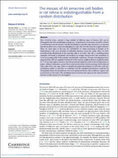
The mosaic of AII amacrine cell bodies in rat retina is indistinguishable from a random distribution
| dc.contributor.author | Liu, Jian Hao | |
| dc.contributor.author | Peter, David Olukoya | |
| dc.contributor.author | Guttormsen, Maren Sofie Faldalen | |
| dc.contributor.author | Hossain, Md Kaykobad | |
| dc.contributor.author | Gerking, Yola | |
| dc.contributor.author | Veruki, Margaret Lin | |
| dc.contributor.author | Hartveit, Espen | |
| dc.date.accessioned | 2022-05-30T09:21:48Z | |
| dc.date.available | 2022-05-30T09:21:48Z | |
| dc.date.created | 2022-05-10T09:41:23Z | |
| dc.date.issued | 2022 | |
| dc.identifier.issn | 0952-5238 | |
| dc.identifier.uri | https://hdl.handle.net/11250/2996708 | |
| dc.description.abstract | The vertebrate retina contains a large number of different types of neurons that can be distinguished by their morphological properties. Assuming that no location should be without a contribution from the circuitry and function linked to a specific type of neuron, it is expected that the dendritic trees of neurons belonging to a type will cover the retina in a regular manner. Thus, for most types of neurons, the contribution to visual processing is thought to be independent of the exact location of individual neurons across the retina. Here, we have investigated the distribution of AII amacrine cells in rat retina. The AII is a multifunctional amacrine cell found in mammals and involved in synaptic microcircuits that contribute to visual processing under both scotopic and photopic conditions. Previous investigations have suggested that AIIs are regularly distributed, with a nearest-neighbor distance regularity index of ~4. It has been argued, however, that this presumed regularity results from treating somas as points, without taking into account their actual spatial extent which constrains the location of other cells of the same type. When we simulated random distributions of cell bodies with size and density similar to real AIIs, we confirmed that the simulated distributions could not be distinguished from the distributions observed experimentally for AIIs in different regions and eccentricities of the retina. The developmental mechanisms that generate the observed distributions of AIIs remain to be investigated. | en_US |
| dc.language.iso | eng | en_US |
| dc.publisher | Cambridge University Press | en_US |
| dc.rights | Navngivelse 4.0 Internasjonal | * |
| dc.rights.uri | http://creativecommons.org/licenses/by/4.0/deed.no | * |
| dc.subject | Neurovitenskap / nevrovitenskap | en_US |
| dc.subject | Neurosciences | en_US |
| dc.subject | Visual neuroscience | en_US |
| dc.subject | Visual neuroscience | en_US |
| dc.title | The mosaic of AII amacrine cell bodies in rat retina is indistinguishable from a random distribution | en_US |
| dc.title.alternative | The mosaic of AII amacrine cell bodies in rat retina is indistinguishable from a random distribution | en_US |
| dc.type | Journal article | en_US |
| dc.type | Peer reviewed | en_US |
| dc.description.version | publishedVersion | en_US |
| dc.rights.holder | Copyright The Author(s), 2022 | en_US |
| dc.source.articlenumber | E004 | en_US |
| cristin.ispublished | true | |
| cristin.fulltext | original | |
| cristin.qualitycode | 1 | |
| dc.identifier.doi | https://doi.org/10.1017/S0952523822000025 | |
| dc.identifier.cristin | 2022943 | |
| dc.source.journal | Visual Neuroscience | en_US |
| dc.relation.project | Norges forskningsråd: 261914 | en_US |
| dc.subject.nsi | VDP::Basale medisinske, odontologiske og veterinærmedisinske fag: 710 | en_US |
| dc.subject.nsi | VDP::Basic medical, dental and veterinary sciences: 710 | en_US |
| dc.identifier.citation | Visual Neuroscience. 2022, 39, E004. | en_US |
| dc.source.volume | 39 | en_US |
Tilhørende fil(er)
Denne innførselen finnes i følgende samling(er)
-
Department of Biomedicine [710]
-
Registrations from Cristin [9791]

Med mindre annet er angitt, så er denne innførselen lisensiert som Navngivelse 4.0 Internasjonal