Topography and clinical features of iris melanoma
| dc.contributor.author | Krohn, Jørgen Gitlesen | |
| dc.contributor.author | Sundal, Kristoffer Våge | |
| dc.contributor.author | Frøystein, Torbjørn | |
| dc.date.accessioned | 2022-06-07T08:00:20Z | |
| dc.date.available | 2022-06-07T08:00:20Z | |
| dc.date.created | 2022-05-09T15:13:15Z | |
| dc.date.issued | 2022 | |
| dc.identifier.issn | 1471-2415 | |
| dc.identifier.uri | https://hdl.handle.net/11250/2997614 | |
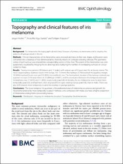
| dc.description.abstract | Background To characterise the topographical and clinical features of primary iris melanoma and to visualise the patterns of tumour extent in the iris. Methods Clinical characteristics of iris melanomas were analysed, and data on their size, shape, and location were converted into a database of two-dimensional iris charts by means of computer-drawing software. The geometric centre of each tumour was entered into corresponding sectors of the chart. The extent of the melanomas was computationally visualised by merging the iris drawings and displaying the number of overlapping tumours on colour-coded iris maps. Results Twenty-nine patients (18 females and 11 males) with a mean age of 52 years met the inclusion criteria. The mean largest tumour diameter was 6.1 mm (range, 1.8–11.0 mm). Five tumours (17%) involved the pupillary margin, 10 (34%) involved the iris root, and 10 (34%) involved both sites. The hemispheric location of the tumour centroid was superior in 3 eyes (11%) and inferior in 25 (89%) (p < 0.0001), and the distribution between the temporal and nasal hemispheres was 17 (61%) and 11 (39%), respectively (p = 0.26). In females, the iris melanomas were located more temporally (p = 0.02) and had more often originated from a pre-existing naevus (p = 0.03), than in males. There was also shift towards more temporally located melanomas in younger patients. Conclusions The lower temporal iris quadrant is the preferential area of melanoma occurrence and growth. Iris melanoma tends to be more temporally located in females, who compared with males also have a higher proportion of melanomas arising from a pre-existing naevus. | en_US |
| dc.language.iso | eng | en_US |
| dc.publisher | BioMed Central | en_US |
| dc.rights | Navngivelse 4.0 Internasjonal | * |
| dc.rights.uri | http://creativecommons.org/licenses/by/4.0/deed.no | * |
| dc.title | Topography and clinical features of iris melanoma | en_US |
| dc.type | Journal article | en_US |
| dc.type | Peer reviewed | en_US |
| dc.description.version | publishedVersion | en_US |
| dc.rights.holder | Copyright 2021 The Author(s) | en_US |
| dc.source.articlenumber | 6 | en_US |
| cristin.ispublished | true | |
| cristin.fulltext | original | |
| cristin.qualitycode | 1 | |
| dc.identifier.doi | 10.1186/s12886-021-02236-3 | |
| dc.identifier.cristin | 2022808 | |
| dc.source.journal | BMC Ophthalmology | en_US |
| dc.identifier.citation | BMC Ophthalmology. 2022, 22 (1), 6. | en_US |
| dc.source.volume | 22 | en_US |
| dc.source.issue | 1 | en_US |
Tilhørende fil(er)
Denne innførselen finnes i følgende samling(er)
-
Department of Clinical Medicine [2095]
-
Registrations from Cristin [10402]

Med mindre annet er angitt, så er denne innførselen lisensiert som Navngivelse 4.0 Internasjonal