Formulation and characterisation of drug-loaded antibubbles for image-guided and ultrasound-triggered drug delivery
| dc.contributor.author | Kotopoulis, Spiros | |
| dc.contributor.author | Lam, Christina | |
| dc.contributor.author | Haugse, Ragnhild | |
| dc.contributor.author | Snipstad, Sofie | |
| dc.contributor.author | Murvold, Elsa Thodesen | |
| dc.contributor.author | Jouleh, Tæraneh | |
| dc.contributor.author | Berg, Sigrid | |
| dc.contributor.author | Hansen, Rune | |
| dc.contributor.author | Popa, Mihaela-Lucia | |
| dc.contributor.author | Mccormack, Emmet Matin | |
| dc.contributor.author | Gilja, Odd Helge | |
| dc.contributor.author | Poortinga, Albert | |
| dc.date.accessioned | 2022-09-14T12:09:24Z | |
| dc.date.available | 2022-09-14T12:09:24Z | |
| dc.date.created | 2022-04-20T10:05:55Z | |
| dc.date.issued | 2022 | |
| dc.identifier.issn | 1350-4177 | |
| dc.identifier.uri | https://hdl.handle.net/11250/3017827 | |
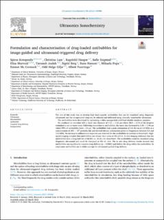
| dc.description.abstract | The aim of this study was to develop high load-capacity antibubbles that can be visualized using diagnostic ultrasound and the encapsulated drug can be released and delivered using clinically translatable ultrasound. The antibubbles were developed by optimising a silica nanoparticle stabilised double emulsion template. We produced an emulsion with a mean size diameter of 4.23 ± 1.63 µm where 38.9 ± 3.1% of the droplets contained a one or more cores. Following conversion to antibubbles, the mean size decreased to 2.96 ± 1.94 µm where 99% of antibubbles were <10 µm. The antibubbles had a peak attenuation of 4.8 dB/cm at 3.0 MHz at a concentration of 200 × 103 particles/mL and showed distinct attenuation spikes at frequencies between 5.5 and 13.5 MHz. No increase in subharmonic response was observed for the antibubbles in contrast to SonoVue®. High-speed imaging revealed that antibubbles can release their cores at MIs of 0.6. In vivo imaging indicated that the antibubbles have a long half-life of 68.49 s vs. 40.02 s for SonoVue®. The antibubbles could be visualised using diagnostic ultrasound and could be disrupted at MIs of ≥0.6. The in vitro drug delivery results showed that antibubbles can significantly improve drug delivery (p < 0.0001) and deliver the drug within the antibubbles. In conclusion antibubbles are a viable concept for ultrasound guided drug delivery. | en_US |
| dc.language.iso | eng | en_US |
| dc.publisher | Elsevier | en_US |
| dc.rights | Navngivelse 4.0 Internasjonal | * |
| dc.rights.uri | http://creativecommons.org/licenses/by/4.0/deed.no | * |
| dc.title | Formulation and characterisation of drug-loaded antibubbles for image-guided and ultrasound-triggered drug delivery | en_US |
| dc.type | Journal article | en_US |
| dc.type | Peer reviewed | en_US |
| dc.description.version | publishedVersion | en_US |
| dc.rights.holder | Copyright 2022 The Author(s) | en_US |
| dc.source.articlenumber | 105986 | en_US |
| cristin.ispublished | true | |
| cristin.fulltext | original | |
| cristin.qualitycode | 1 | |
| dc.identifier.doi | 10.1016/j.ultsonch.2022.105986 | |
| dc.identifier.cristin | 2017772 | |
| dc.source.journal | Ultrasonics Sonochemistry | en_US |
| dc.identifier.citation | Ultrasonics Sonochemistry. 2022, 85, 105986. | en_US |
| dc.source.volume | 85 | en_US |
Files in this item
This item appears in the following Collection(s)
-
Department of Clinical Medicine [2044]
-
Registrations from Cristin [9489]

Except where otherwise noted, this item's license is described as Navngivelse 4.0 Internasjonal