What MRI-based tumor size measurement is best for predicting long-term survival in uterine cervical cancer?
| dc.contributor.author | Lura, Njål Gjærde | |
| dc.contributor.author | Wagner-Larsen, Kari Strøno | |
| dc.contributor.author | Forsse, David Erik | |
| dc.contributor.author | Trovik, Jone | |
| dc.contributor.author | Halle, Mari Kyllesø | |
| dc.contributor.author | Bertelsen, Bjørn | |
| dc.contributor.author | Salvesen, Øyvind | |
| dc.contributor.author | Woie, Kathrine | |
| dc.contributor.author | Krakstad, Camilla | |
| dc.contributor.author | Haldorsen, Ingfrid S. | |
| dc.date.accessioned | 2022-09-22T10:26:14Z | |
| dc.date.available | 2022-09-22T10:26:14Z | |
| dc.date.created | 2022-09-16T13:08:50Z | |
| dc.date.issued | 2022-06-17 | |
| dc.identifier.issn | 1869-4101 | |
| dc.identifier.uri | https://hdl.handle.net/11250/3020633 | |
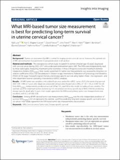
| dc.description.abstract | Background: Tumor size assessment by MRI is central for staging uterine cervical cancer. However, the optimal role of MRI-derived tumor measurements for prognostication is still unclear. Material and methods: This retrospective cohort study included 416 women (median age: 43 years) diagnosed with cervical cancer during 2002–2017 who underwent pretreatment pelvic MRI. The MRIs were independently read by three radiologists, measuring maximum tumor diameters in three orthogonal planes and maximum diameter irrespective of plane (MAXimaging). Inter-reader agreement for tumor size measurements was assessed by intraclass correlation coefficients (ICCs). Size was analyzed in relation to age, International Federation of Gynecology and Obstetrics (FIGO) (2018) stage, histopathological markers, and disease-specific survival using Kaplan–Meier-, Cox regression-, and time-dependent receiver operating characteristics (tdROC) analyses. Results: All MRI tumor size variables (cm) yielded high areas under the tdROC curves (AUCs) for predicting survival (AUC 0.81–0.84) at 5 years after diagnosis and predicted outcome (hazard ratios [HRs] of 1.42–1.76, p < 0.001 for all). Only MAXimaging independently predicted survival (HR = 1.51, p = 0.03) in the model including all size variables. The optimal cutoff for maximum tumor diameter (≥ 4.0 cm) yielded sensitivity (specificity) of 83% (73%) for predicting disease-specific death after 5 years. Inter-reader agreement for MRI-based primary tumor size measurements was excellent, with ICCs of 0.83–0.85. Conclusion: Among all MRI-derived tumor size measurements, MAXimaging was the only independent predictor of survival. MAXimaging ≥ 4.0 cm represents the optimal cutoff for predicting long-term disease-specific survival in cervical cancer. Inter-reader agreement for MRI-based tumor size measurements was excellent. | en_US |
| dc.language.iso | eng | en_US |
| dc.publisher | Springer Open | en_US |
| dc.rights | Navngivelse 4.0 Internasjonal | * |
| dc.rights.uri | http://creativecommons.org/licenses/by/4.0/deed.no | * |
| dc.title | What MRI-based tumor size measurement is best for predicting long-term survival in uterine cervical cancer? | en_US |
| dc.type | Journal article | en_US |
| dc.type | Peer reviewed | en_US |
| dc.description.version | publishedVersion | en_US |
| dc.rights.holder | Copyright 2022 the authors | en_US |
| dc.source.articlenumber | 105 | en_US |
| cristin.ispublished | true | |
| cristin.fulltext | original | |
| cristin.qualitycode | 1 | |
| dc.identifier.doi | 10.1186/s13244-022-01239-y | |
| dc.identifier.cristin | 2052484 | |
| dc.source.journal | Insight into Imaging | en_US |
| dc.identifier.citation | Insight into Imaging. 2022, 13, 105. | en_US |
| dc.source.volume | 13 | en_US |
Tilhørende fil(er)
Denne innførselen finnes i følgende samling(er)
-
Department of Clinical Medicine [2066]
-
Registrations from Cristin [9791]

Med mindre annet er angitt, så er denne innførselen lisensiert som Navngivelse 4.0 Internasjonal