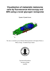
Visualization of metastatic melanoma cells by fluorescence microscopy and MRI using a novel glycogen nanoprobe
Master thesis
Permanent lenke
https://hdl.handle.net/1956/8228Utgivelsesdato
2014-06-05Metadata
Vis full innførselSamlinger
Sammendrag
The incidence of malignant melanoma has increased steadily during the last decades. Large portions of individuals with this particular skin cancer develop multiple brain metastases, which is associated with a particular poor prognosis, and thus new therapeutic approaches are needed. Increased attention has been given lately in literature to the establishment of functional nano-scaled materials for applications in combined cancer therapy and diagnostics, a field termed «theranostics» or «theragnosis» (therapy + diagnostics). Recently, a collaborator at the Institute of Macromolecular Chemistry (Academy of Sciences, Prague) developed a multimodal nanoprobe, consisting of a backbone of glycogen. Two different contrast agents were encompassed in this, namely Dyomics-615-NHS ester, a fluorescent marker and Gd-DOTA, a well-known magnetic resonance imaging contrast agent. The nanoprobe can also be loaded with positron emission tomography tracers and therapeutic substances, and targeting may be achieved by loading the probe with antibodies. The nanoprobe has a great potential for being a theranostic probe, as the backbone consists of glycogen, which is regarded nontoxic to human cells. Further, magnetic resonance imaging and positron emission tomography may be used to evaluate drug uptake and treatment effects. Since this nanoprobe is completely new, it has not been tested previosly on human cell lines in vitro and in vivo. The main aim of the current Master thesis was thus to perform toxicity and viability studies in vitro, to determine the usefulness of the nanoprobe. The usability of the nanoprobe was evaluated on three different metastatic melanoma cell lines in vitro. Fluorescence microscopy and high throughput imaging revealed a high cell labeling efficacy with an optimal uptake period at 24 hours incubation time. Reduced cell viability was not found after labeling with the nanoprobe. In vitro magnetic resonance imaging studies of labeled cells casted in agar phantoms revealed that the nanoprobe also can be used as a contrast agent by this modality. The preliminary in vivo data indicated that tumor contrast uptake could be achieved in a subcutaneous tumor model. The promising results reported in this thesis may indicate that this nanoprobe can be used also for other cancer cell lines. Potentially, the nanoprobe can offer further benefits over established contrast agents used in magnetic resonance imaging, as pharmaceuticals attached to the probe can be traced simultaneously as the progression of disease and treatment are monitored.